Pelvic Anatomy Female Ligaments - Anatomical Teaching Models - Plastic Human Pelvic Models ... / The pelvis is held together by three principal ligaments:
byAdmin-
0
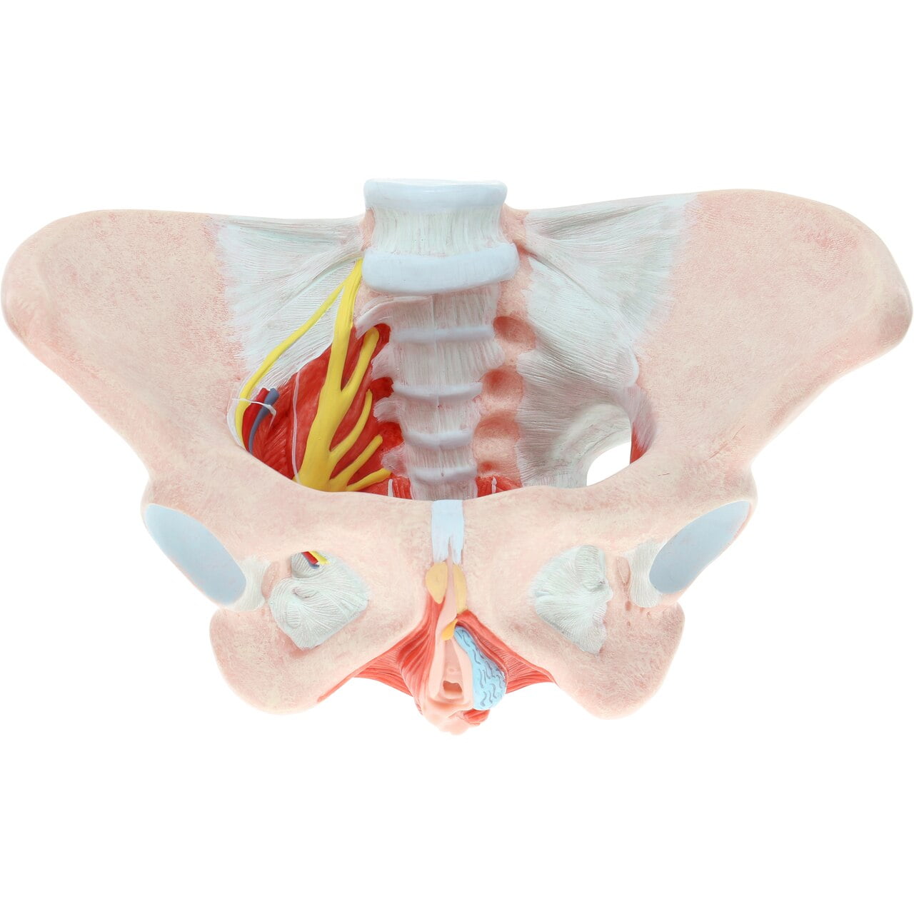
Pelvic Anatomy Female Ligaments - Anatomical Teaching Models - Plastic Human Pelvic Models ... / The pelvis is held together by three principal ligaments:. This occurs because of weaknesses in pelvic support mechanisms (i.e., pelvic ligaments) that sometimes are associated with repeated stretching due to multiple pregnancies. This image shows the posterior back view of the female pelvic brim (the bones and ligaments that forms the pelvic region in the female) showing: The outlet is formed by the pubic arch, ischial spines, sacrotuberous ligaments, and the coccyx. Pelvic anatomy sacrouterine ligament cardinal ligaments pelvic fascia sacrospinous ligament urethral support bladder support rectal support. Finally, a checklist is provided for structured reporting of the mri findings in the female pelvis.
These ligaments also play a crucial role in pelvic organ prolapse with anterior vaginal wall descent (5). Additional ligaments may be found in the female pelvis. Sagittal t2 mr image of the pelvis Functional anatomy of the male pelvicfloor explore the important aspects of the structures and functions of the male pelvic. If you are looking for an anatomy mo
Axis Scientific Female Pelvic Floor with Muscles ... from i5.walmartimages.com Imaios and selected third parties, use cookies or similar technologies, in particular for audience measurement. Also, bladder function problems after spinal cord injury are discussed. It provides attachment to some important muscles in the region, and forms a cavity which accommodates several important internal organs. These ligaments are important stabilizers. The right half shows the bones with pelvic ligaments. It extends to both sides of the pelvic wall. Finally, a checklist is provided for structured reporting of the mri findings in the female pelvis. Female pelvis with ligaments muscles and organs, 4 part.
The broad ligament can be further divided into three components that are linked to.
Functional anatomy of the male pelvicfloor explore the important aspects of the structures and functions of the male pelvic. They're blends of fascias and other ligaments that form an independent structure. • lateral boundaries—fused ilium and ischium. The relationship of the pelvic floor and the organs exiting the pelvis is shown above. This anatomically detailed model is a great way to teach and learn the anatomy of the human female pelvis. It is strengthened and supported by several joints and ligaments. Other ligaments attached to bony pelvis include the sacrococcygeal ligaments, pubic symphysis ligaments, and endopelvic fascia ligament. Of female pelvic organ sacrospinous ligament just medial to the ischial spine, exiting the pelvis through the greater sciatic foramen. If you are looking for an anatomy mo Finally, a checklist is provided for structured reporting of the mri findings in the female pelvis. • muscles and ligaments form a pelvic floor. The ligaments of the female reproductive tract can be divided into three categories: The broad ligament of the uterus serves as mesentery to the female pelvic organs and contains blood vessels, nerves, and lymphatics.
Obviously, some ligaments are found in the female pelvis but not the male. The cardinal and uterosacral ligaments, which support the uterus and upper part of the vagina, are critical structures in the female pelvis (3, 4). See ligaments of the female pelvis below. Also, bladder function problems after spinal cord injury are discussed. • posterolateral wall—piriformis and coccygeus muscles.
Female Pelvis Model with Ligaments, Vessels, Nerves and ... from www.anatomystuff.co.uk The right half shows the bones with pelvic ligaments. The relationship of the pelvic floor and the organs exiting the pelvis is shown above. Cookies allow us to analyze and store information such as the characteristics of your device as well as certain personal data (e.g., ip addresses, navigation, usage or geolocation data, unique identifiers). Iliolumbar, sacrotuberous and sacrospinous ligaments. It is strengthened and supported by several joints and ligaments. • located inferior to the pelvic brim. The pelvis is held together by three principal ligaments: The ligaments of the female reproductive tract can be divided into three categories:
• located inferior to the pelvic brim.
Obviously, some ligaments are found in the female pelvis but not the male. The broad ligament can be further divided into three components that are linked to. It provides attachment to some important muscles in the region, and forms a cavity which accommodates several important internal organs. • lateral boundaries—fused ilium and ischium. They're blends of fascias and other ligaments that form an independent structure. The pelvis is a basin shaped bony structure formed by the combination of two pelvic bones (hip bones or innominate bones) and the sacrum. Finally, a checklist is provided for structured reporting of the mri findings in the female pelvis. Pelvic anatomy sacrouterine ligament cardinal ligaments pelvic fascia sacrospinous ligament urethral support bladder support rectal support. Ligaments connect one bone to another and provide important stability. Pelvic surgery requires a comprehensive knowledge of the pelvic anatomy to safely attain access, maximize exposure, ensure hemostasis, and avoid injury to viscera, blood vessels, and nerves. The broad ligament of the uterus serves as mesentery to the female pelvic organs and contains blood vessels, nerves, and lymphatics. Many pelvic landmarks, ligaments, and muscular structures within the pelvis are important to know to differentiate normal reproductive organs from muscular and vascular structures. This image shows the posterior back view of the female pelvic brim (the bones and ligaments that forms the pelvic region in the female) showing:
See ligaments of the female pelvis below. Other ligaments attached to bony pelvis include the sacrococcygeal ligaments, pubic symphysis ligaments, and endopelvic fascia ligament. A collection of anatomy notes covering the key anatomy concepts that medical students need to learn. The cardinal and uterosacral ligaments, which support the uterus and upper part of the vagina, are critical structures in the female pelvis (3, 4). It is strengthened and supported by several joints and ligaments.
Module 5: Pelvis Imaging from www.hitachihealthcare.com This section of the website will explain large and minute details of axial male pelvis cross sectional anatomy. Additional ligaments may be found in the female pelvis. • posterolateral wall—piriformis and coccygeus muscles. • lateral boundaries—fused ilium and ischium. Sagittal t2 mr image of the pelvis A sagittal view of the female pelvis is shown in the figure ( figure 1 ). If you are looking for an anatomy mo Obviously, some ligaments are found in the female pelvis but not the male.
The inlet to the pelvic canal is at the level of the sacral promontory and superior aspect of the pubic bones.
Imaios and selected third parties, use cookies or similar technologies, in particular for audience measurement. Female pelvis with ligaments muscles and organs, 4 part. • muscles and ligaments form a pelvic floor. • posterolateral wall—piriformis and coccygeus muscles. Interactive video showing normal female pelvic anatomy as seen by laparoscopy. The outlet is formed by the pubic arch, ischial spines, sacrotuberous ligaments, and the coccyx. Female pelvis model with ligaments, muscles & organs h20/3. The broad ligament can be further divided into three components that are linked to. The ligaments of the female reproductive tract can be divided into three categories: Functional anatomy of the pelvic floor. A sagittal view of the female pelvis is shown in the figure ( figure 1 ). The pelvis is held together by three principal ligaments: A collection of anatomy notes covering the key anatomy concepts that medical students need to learn.
(see surgical female urogenital anatomy, section on 'lower genital pelvic anatomy. The inlet to the pelvic canal is at the level of the sacral promontory and superior aspect of the pubic bones.